| dc.contributor.author | Løken, Sverre | |
| dc.contributor.author | Ludvigsen, Tom C. | |
| dc.contributor.author | Høysveen, Turid | |
| dc.contributor.author | Holm, Inger | |
| dc.contributor.author | Engebretsen, Lars | |
| dc.contributor.author | Reinholt, Finn P. | |
| dc.date.accessioned | 2010-07-07T12:27:42Z | |
| dc.date.available | 2010-07-07T12:27:42Z | |
| dc.date.issued | 2009-07-02 | |
| dc.identifier | Seksjon for idrettsmedisinske fag / Department of Sports Medicine | |
| dc.identifier.citation | Knee Surgery, Sports Traumatology, Arthroscopy. 2009, 17(11), 1278-1288 | en_US |
| dc.identifier.issn | 0942-2056 | |
| dc.identifier.uri | http://hdl.handle.net/11250/170623 | |
| dc.description | I Brage finner du siste tekst-versjon av artikkelen, og den kan inneholde ubetydelige forskjeller fra forlagets pdf-versjon. Forlagets pdf-versjon finner du på www.springerlink.com: http://dx.doi.org/10.1007/s00167-009-0854-5 / In Brage you'll find the final text version of the article, and it may contain insignificant differences from the journal's pdf version. The original publication is available at www.springerlink.com: http://dx.doi.org/10.1007/s00167-009-0854-5 | en_US |
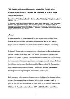
| dc.description.abstract | Autologous chondrocyte implantation (ACI)
usually results in improvement in clinical scores. However,
long-term isokinetic muscle strength measurements have
not been reported. Biopsies from the repair tissue have
shown variable proportions of hyaline-like cartilage. In this
study, 21 consecutive patients were treated with autologous
cartilage implantations in the knee. Mean size of the lesions
was 5.5 cm2. Follow-up arthroscopy with biopsy was performed
at 2 years in 19 patients. The biopsies were examined
with both light microscopy and transmission electron
microscopy (TEM) techniques including immunogold
analysis of collagen type 1. Patient function was evaluated
with modified 10-point scales of the Cincinnati knee rating
system obtained preoperatively and at 1 and 8.1 years.
Isokinetic quadriceps and hamstrings muscle strength testing
was performed at 1, 2 and 7.4 years. Light microscopy
and TEM both showed predominately fibrous cartilage. The
immunogold analysis showed a high percentage of collagen
type I. At 7.4 years, the total work deficits when compared
with the contra-lateral leg for isokinetic extension were 19.1
and 11.4%, and for isokinetic flexion 11.8 and 8.5% for 60
and 2408/s, respectively. Mean pain score improved from
4.3 preoperatively to 6.3 at 1 year (p = 0.031) and 6.6 at
8.1 years (p = 0.013). Overall health condition score
improved from 4.1 preoperatively to 6.1 at 1 year
(p = 0.004) and 6.5 at 8.1 years (p = 0.008). Three
patients later went through revision surgery with other
resurfacing techniques and are considered failures. In
summary, the formation of fibrous cartilage following ACI
was confirmed by TEM with immunogold histochemistry.
Although the functional scores were generally good,
strength measurements demonstrated that the surgically
treated leg remained significantly weaker. | en_US |
| dc.language.iso | eng | en_US |
| dc.publisher | Springer | en_US |
| dc.subject | articular cartilage | en_US |
| dc.subject | chondrocytes | en_US |
| dc.subject | autologous chondrocyte implantation | en_US |
| dc.subject | muscle strength | en_US |
| dc.subject | surgery | en_US |
| dc.subject | knee | en_US |
| dc.subject | histology | en_US |
| dc.subject | microscopy | en_US |
| dc.subject | electron | en_US |
| dc.title | Autologous chondrocyte implantation to repair knee cartilage injury : ultrastructural evaluation at 2 years and long term follow up including muscle strength measurements | en_US |
| dc.type | Journal article | en_US |
| dc.type | Peer reviewed | en_US |
| dc.subject.nsi | VDP::Medical disciplines: 700 | en_US |
| dc.source.pagenumber | 1278-1288 | en_US |