| dc.contributor.author | Forså, Marianne Inngjerdingen | |
| dc.contributor.author | Smedsrud, Marit Kristine | |
| dc.contributor.author | Haugaa, Kristina Ingrid Helena Hermann | |
| dc.contributor.author | Bjerring, Anders W. | |
| dc.contributor.author | Fruh, Andreas | |
| dc.contributor.author | Sarvari, Sebastian I. | |
| dc.contributor.author | Landgraff, Hege Elisabeth Wilson | |
| dc.contributor.author | Hallén, Jostein | |
| dc.contributor.author | Edvardsen, Thor | |
| dc.date.accessioned | 2024-03-18T13:34:08Z | |
| dc.date.available | 2024-03-18T13:34:08Z | |
| dc.date.created | 2024-02-15T08:27:35Z | |
| dc.date.issued | 2023 | |
| dc.identifier.citation | European Journal of Preventive Cardiology. 2023, Artikkel zwad361. | en_US |
| dc.identifier.issn | 2047-4873 | |
| dc.identifier.uri | https://hdl.handle.net/11250/3122923 | |
| dc.description | This is an Open Access article distributed under the terms of the Creative Commons Attribution-NonCommercial License (https://creativecommons.org/licenses/by-nc/4.0/), which permits non-commercial re-use, distribution, and reproduction in any medium, provided the original work is properly cited. | en_US |
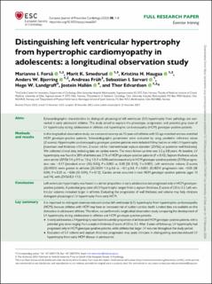
| dc.description.abstract | Aims: Echocardiographic characteristics to distinguish physiological left ventricular (LV) hypertrophy from pathology are warranted in early adolescent athletes. This study aimed to explore the phenotype, progression, and potential grey zone of LV hypertrophy during adolescence in athletes and hypertrophic cardiomyopathy (HCM) genotype–positive patients.
Methods and results: In this longitudinal observation study, we compared seventy-six 12-year-old athletes with 55 age-matched and sex-matched HCM genotype–positive patients. Echocardiographic parameters were evaluated by using paediatric reference values (Z-scores). Hypertrophic cardiomyopathy genotype–positive patients were included if they had no or mild LV hypertrophy [maximum wall thickness <13 mm, Z-score <6 for interventricular septum diameter (ZIVSd), or posterior wall thickness]. We collected clinical data, including data on cardiac events. The mean follow-up-time was 3.2 ± 0.8 years. At baseline, LV hypertrophy was found in 28% of athletes and 21% of HCM genotype–positive patients (P = 0.42). Septum thickness values were similar (ZIVSd 1.4 ± 0.9 vs. 1.0 ± 1.3, P = 0.08) and increased only in HCM genotype–positive patients {ZIVSd progression rate −0.17 [standard error (SE) 0.05], P = 0.002 vs. 0.30 [SE 0.10], P = 0.001}. Left ventricular volume Z-scores (ZLVEDV) were greater in athletes [ZLVEDV 1.0 ± 0.6 vs. −0.1 ± 0.8, P < 0.001; ZLVEDV progression rate −0.05 (SE 0.04), P = 0.21 vs. −0.06 (SE 0.04), P = 0.12]. Cardiac arrest occurred in two HCM genotype–positive patients (ages 13 and 14), with ZIVSd 8.2–11.5.
Conclusion: Left ventricular hypertrophy was found in a similar proportion in early adolescence but progressed only in HCM genotype–positive patients. A potential grey zone of LV hypertrophy ranged from a septum thickness Z-score of 2.0 to 3.3. Left ventricular volumes remained larger in athletes. Evaluating the progression of wall thickness and volume may help clinicians distinguish physiological LV hypertrophy from early HCM. | en_US |
| dc.language.iso | eng | en_US |
| dc.subject | adolescent | en_US |
| dc.subject | athlete | en_US |
| dc.subject | cardiac remodelling | en_US |
| dc.subject | echocardiography | en_US |
| dc.title | Distinguishing left ventricular hypertrophy from hypertrophic cardiomyopathy in adolescents: A longitudinal observation study | en_US |
| dc.type | Peer reviewed | en_US |
| dc.type | Journal article | en_US |
| dc.description.version | publishedVersion | en_US |
| dc.rights.holder | © The Author(s) 2023 | en_US |
| dc.source.pagenumber | 8 | en_US |
| dc.source.journal | European Journal of Preventive Cardiology | en_US |
| dc.identifier.doi | 10.1093/eurjpc/zwad361 | |
| dc.identifier.cristin | 2246206 | |
| dc.description.localcode | Institutt for fysisk prestasjonsevne / Department of Physical Performance | en_US |
| dc.source.articlenumber | zwad361 | en_US |
| cristin.ispublished | true | |
| cristin.fulltext | original | |
| cristin.qualitycode | 2 | |
